Diagram Of Bones In Neck And Shoulder / Labeled Anatomy Chart Of Shoulder Ligaments On White Background Stock Photo Download Image Now Istock - Here is an overview of the shoulder bones:
Diagram Of Bones In Neck And Shoulder / Labeled Anatomy Chart Of Shoulder Ligaments On White Background Stock Photo Download Image Now Istock - Here is an overview of the shoulder bones:. Bone diagram forehead (frontal bone) nose bones (nasals) cheek bone (zygoma) upper jaw (maxilla) lower jaw (mandible) breast bone (sternum) upper arm bone (humerus) lower arm bone (ulna) thigh bone (femur) collar bone (clavicle) toe bones (phalanges) ankle bones (tarsals) kneecap (patella) shin bone (tibia) calf bone (fibula) foot bones It is made up of bones, discs, muscles, ligaments, nerves and tendons. I was operated 3 years ago from my neck and got 3 bones removed it is the most painful feeling a human will ever experience i now have titanium plates&roades w/ nummness on both arm& secure pain for the rest of my life ,but i had been told that i had week bones as a child but i dont even wish this on my worst enemy/ if you have any type of pain on your neck please & i mean please get it check. The column of the neck bones is slightly curved. Related posts of diagram of shoulder muscles and tendons anatomy muscles view.
Bone diagram forehead (frontal bone) nose bones (nasals) cheek bone (zygoma) upper jaw (maxilla) lower jaw (mandible) breast bone (sternum) upper arm bone (humerus) lower arm bone (ulna) thigh bone (femur) collar bone (clavicle) toe bones (phalanges) ankle bones (tarsals) kneecap (patella) shin bone (tibia) calf bone (fibula) foot bones A second joint in the shoulder is the junction of the collar bone with the shoulder blade, called the acromioclavicular joint. Shoulder dislocations and surgical neck fractures (breaks) of the humerus can therefore be accompanied by nerve injury. It is commonly referred to as the shoulder blade. Neck and shoulder muscles diagram neck shoulder muscle anatomy shoulder muscle anatomy diagram anatomy.
:background_color(FFFFFF):format(jpeg)/images/article/en/shoulder-girdle/Uo74gpKOKbev6FDAqJY6w_clavicula_3_large_qcx4M64XQJzhdnOpuqs67Q.png)
Anatomy muscles view 12 photos of the anatomy muscles view anatomy muscles view, anatomy of body muscles back view, muscle anatomy anterior view, muscle anatomy back view, muscle anatomy posterior view, human muscles, anatomy muscles view, anatomy of body muscles back view, muscle anatomy anterior view, muscle.
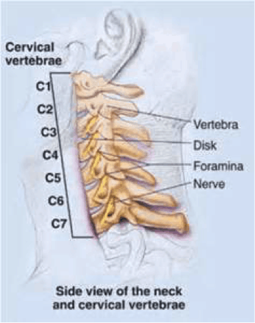
#bones in the head neck and shoulder #bones of the head neck and shoulder girdle #bones of the head neck and shoulders #function of the head neck and shoulder bones #the position of the head face neck chest and shoulder girdle bones The neck is connected to the upper back through a series of seven vertebral segments. Human shoulder anatomy muscles ligaments and… continue reading → Think of it like a jigsaw puzzle, all the pieces fit in together and are required to get the full picture as to how it works. Neck and shoulders muscles diagram. For more anatomy content please follow us and visit our website: The anatomy of the neck and shoulders is very interesting. Diagram of human shoulder muscles / shoulder anatomy campbell hand shoulder surgeon southampton : In order to reposition your neck to its optimal neutral alignment, we must first move it gently through a full range of motion. Muscles allow us to move by pulling on bones. Human shoulder muscles and joints have a red signal. Another name for this bone is the shoulder blade. The top of the cervical spine connects to the skull, and the bottom connects to the upper back at about shoulder level.
Identify the bony structures and key landmarks of the neck and shoulder complex. The humerus or one of the other bones in the shoulder slips out of position. Raising the arm causes pain and a popping sensation if the shoulder is dislocated. It connects the humerus (upper arm) with the clavicle. It helps stabilize the shoulder's movements.

The bones of the shoulder are as given below.
The scapula is a flat, triangular bone that forms the posterior part of the shoulder girdle. Here is an overview of the shoulder bones: Anatomy muscles view 12 photos of the anatomy muscles view anatomy muscles view, anatomy of body muscles back view, muscle anatomy anterior view, muscle anatomy back view, muscle anatomy posterior view, human muscles, anatomy muscles view, anatomy of body muscles back view, muscle anatomy anterior view, muscle. The acromion and the glenoid. It is made up of bones, discs, muscles, ligaments, nerves and tendons. The bones of the superior portion of the skull are known as the cranium and protect the brain from damage. Another name for this bone is the shoulder blade. Although anchored in the neck, their primary functions are to move the shoulder blades and support the arms. In order to reposition your neck to its optimal neutral alignment, we must first move it gently through a full range of motion. There are seven of them. Diagram of human shoulder muscles / shoulder anatomy campbell hand shoulder surgeon southampton : The bones of the shoulder are as given below. Think of it like a jigsaw puzzle, all the pieces fit in together and are required to get the full picture as to how it works.
We also prepared a custom quiz on the neck anatomy. Identify the key joint structures of the neck and shoulder region. Human shoulder anatomy muscles ligaments and… continue reading → The first one that holds the skull is called the atlas. Think of it like a jigsaw puzzle, all the pieces fit in together and are required to get the full picture as to how it works.

At the completion of unit 10 the student will be able to:
The anatomy of the neck and shoulders is very interesting. Use a barbell, a pair of dumbbells, or a shoulder press machine and. Here is an overview of the shoulder bones: We are pleased to provide you with the picture named head and neck muscles diagram.we hope this picture head and neck muscles diagram can help you study and research. At the completion of unit 10 the student will be able to: #bones in the head neck and shoulder #bones of the head neck and shoulder girdle #bones of the head neck and shoulders #function of the head neck and shoulder bones #the position of the head face neck chest and shoulder girdle bones The upper arm bone, called the humerus, is connected to the body via the shoulder blade, which possesses the latin name scapula. Raising the arm causes pain and a popping sensation if the shoulder is dislocated. There are anterior muscles diagrams and posterior muscles. For more anatomy content please follow us and visit our website: The humerus or one of the other bones in the shoulder slips out of position. The top of the cervical spine connects to the skull, and the bottom connects to the upper back at about shoulder level. Neck and shoulder muscles diagram neck shoulder muscle anatomy shoulder muscle anatomy diagram anatomy.
Komentar
Posting Komentar